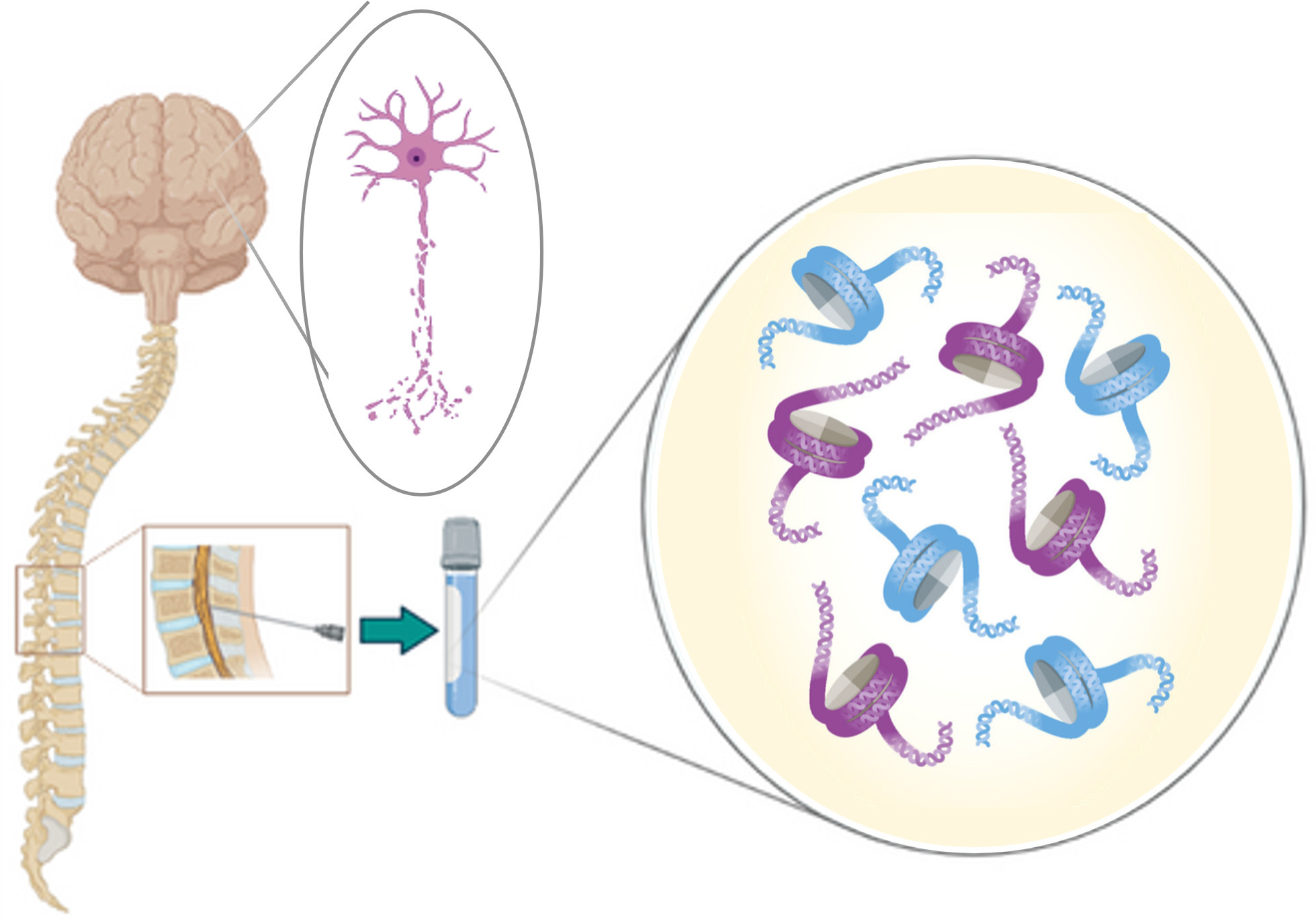
Given the difficulty of tissue sampling the use of circulating biomarkers in neurology is a growing field. Recent diagnostic approaches that use circulating protein biomarkers, such as neurofilament light chain, are gaining traction. However, these biomarkers are applicable to single diseases only and provide limited insight into underlying disease biology. The analysis of cell free DNA (cfDNA) methylation biomarkers provides insight into disease biology through tissue of origin1 (TOO) and gene expression inference2 analysis. Coupled with the observation that 5hmC levels are elevated in neuronal tissues3 there is an opportunity to improve the detection and monitoring of neurological conditions through 6-base analysis of cfDNA derived from cerebrospinal fluid, the most proximal biofluid to the brain.
The Jagodic group at Karolinska Institutet have done just that. They analysed 6-base data derived from CSF cfDNA from healthy individuals and those with Multiple Sclerosis (MS), Traumatic Brain Injury (TBI), Progressive Multifocal Leukoencephalopathy (PML), Amyotrophic Lateral Sclerosis (ALS) and other movement disorders (MovDis).
Despite there being very little cfDNA in CSF (far less than a nanogram, often so little that it’s not detectable with conventional DNA quantification methods) the team prepared and sequenced duet evoC libraries to produce high quality 6-base data. The ability to distinguish 5mC from 5hmC was critical to the objectives of the study, not just because of the potential for 5hmC to provide deeper TOO and mechanistic insight from cfDNA, but also because 5hmC is elevated in neuronal tissues. In fact, the team 6-base sequenced sorted brain cell types and saw exactly that; more than 35% 5hmC in neurons and ~20% 5hmC in glial cells.
Armed with this data the team explored levels of 5mC and 5hmC across cfDNA from the different neurological disorders and were able to distinguish each group, and healthy controls, using multiomic factor analysis. They used 5mC and 5hmC regions known to be relevant to brain biology to build and test a classifier that distinguished healthy controls from disease with a high AUC of 0.936, demonstrating the power of 6-base data to detect neurological disease even where cfDNA levels are so low they cannot be quantified.
Gene set enrichment analysis of regions with elevated 5mC and 5hmC across the cohort revealed disease specific biology; for example there was enrichment of immune signatures in MS and glial cells in MovDis compared to controls. Taking this further, cellular deconvolution approaches demonstrated CD8+ T cell contributions to MS CSF cfDNA and endothelial cells in TBI.
Finally, the team used the modality XPLR software to detect differentially methylated regions (DMRs) in their data. These DMRs revealed impacts on olfactory receptors in TBI, where individuals often lose their sense of smell, and synaptic processes in MS, which reflects the known pathology of the disease.
The team conclude that 6-base data can be used to detect neuronal damage and has the potential for real time investigation and monitoring of neurological conditions.
1. Loyfer, N. et al. A DNA methylation atlas of normal human cell types. Nature 613, 355–364 (2023).
2. Sowalsky, A. et al. Integrative dual ctDNA 5mC/5hmC methylomics and clonal reconstruction infer tumor transcription and resistance phenotypes in metastatic prostate cancer. Preprint at https://doi.org/10.21203/rs.3.rs-8778762/v1 (2026).
3. Kriaucionis, S. & Heintz, N. The nuclear DNA base, 5-hydroxymethylcytosine is present in brain and enriched in Purkinje neurons. Science 324, 929–930 (2009).