- Luke Sarre¹
- Aurelie Modat¹
- Lidia Prieto-Lafuente¹
- Tom Charlesworth¹
- Angela Simeone¹
- Mike Stubbington¹
1 biomodal Ltd, The Trinity Building, Chesterford Research Park, Cambridge, UK
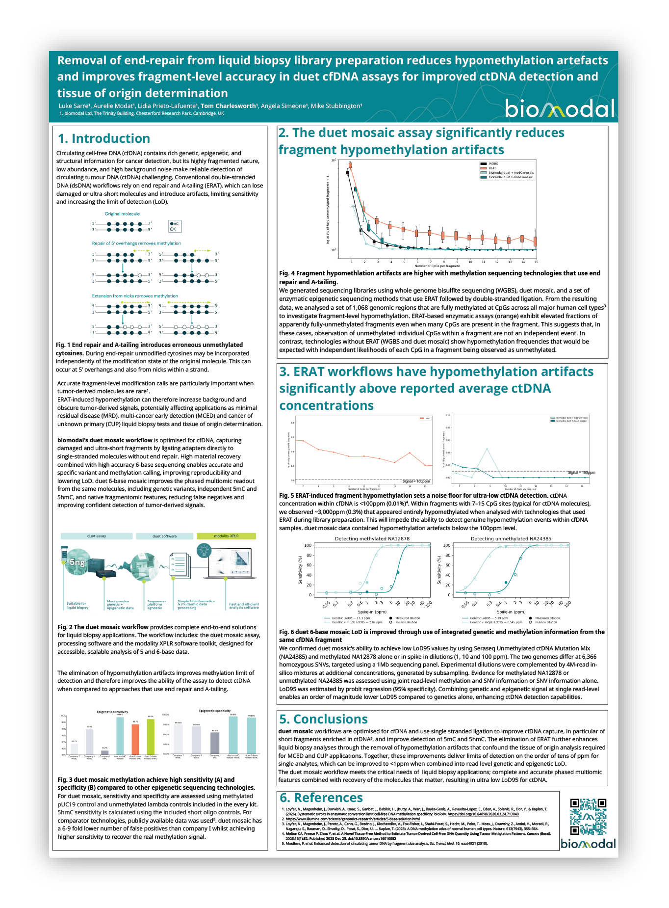
Circulating cell-free DNA (cfDNA) contains rich genetic, epigenetic, and structural information for cancer detection, but its highly fragmented nature, low abundance, and high background noise make reliable detection of circulating tumour DNA (ctDNA) challenging. Conventional double-stranded DNA (dsDNA) workflows rely on end repair and A-tailing (ERAT), which can lose damaged or ultra-short molecules and introduce artifacts, limiting sensitivity and increasing the limit of detection (LoD).
Accurate fragment-level modification calls are particularly important when tumor-derived molecules are rare¹. ERAT-induced hypomethylation can therefore increase background and obscure tumor-derived signals, potentially affecting applications as minimal residual disease (MRD), multi-cancer early detection (MCED) and cancer of unknown primary (CUP) liquid biopsy tests and tissue of origin determination.
biomodal’s duet mosaic workflow is optimised for cfDNA, capturing damaged and ultra-short fragments by ligating adapters directly to single-stranded molecules without end repair. High material recovery combined with high accuracy 6-base sequencing enables accurate and specific variant and methylation calling, improving reproducibility and lowering LoD. duet 6-base mosaic improves the phased multiomic readout from the same molecules, including genetic variants, independent 5mC and 5hmC, and native fragmentomic features, reducing false negatives and improving confident detection of tumor-derived signals.
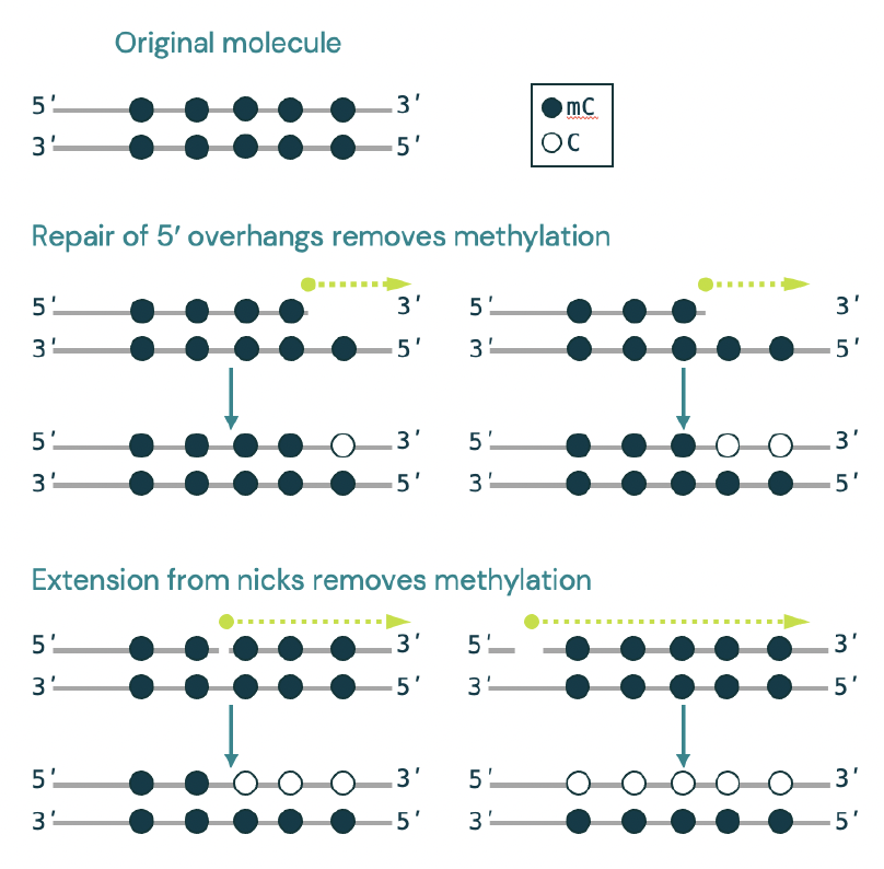
Fig. 1 End repair and A-tailing introduces erroneous unmethylated cytosines. During end-repair unmodified cytosines may be incorporated independently of the modification state of the original molecule. This can occur at 5ʹ overhangs and also from nicks within a strand.
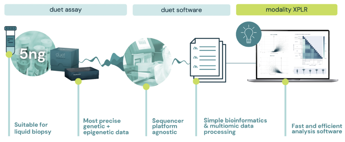
Fig. 2 The duet mosaic workflow provides complete end-to-end solutions for liquid biopsy applications. The workflow includes: the duet mosaic assay, processing software and the modality XPLR software toolkit, designed for accessible, scalable analysis of 5 and 6-base data.
The elimination of hypomethylation artifacts improves methylation limit of detection and therefore improves the ability of the assay to detect ctDNA when compared to approaches that use end repair and A-tailing.
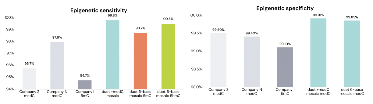
Fig. 3 duet mosaic methylation achieve high sensitivity (A) and specificity (B) compared to other epigenetic sequencing technologies. For duet mosaic, sensitivity and specificity are assessed using methylated pUC19 control and unmethylated lambda controls included in the every kit. 5hmC sensitivity is calculated using the included short oligo controls. For comparator technologies, publicly available data was used². duet mosaic has a 6-9 fold lower number of false positives than company I whilst achieving higher sensitivity to recover the real methylation signal.
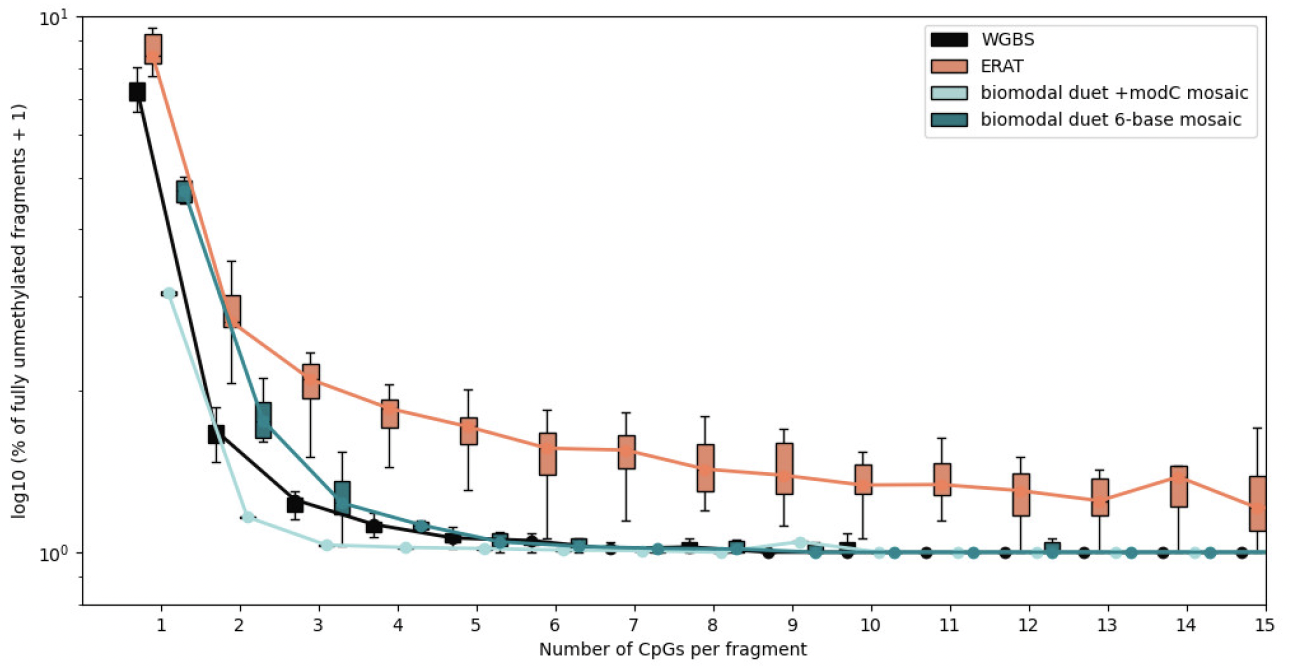
Fig. 4 Fragment hypomethlation artifacts are higher with methylation sequencing technologies that use end repair and A-tailing. We generated sequencing libraries using whole genome bisulfite sequencing (WGBS), duet mosaic, and a set of enzymatic epigenetic sequencing methods that use ERAT followed by double-stranded ligation. From the resulting data, we analysed a set of 1,068 genomic regions that are fully methylated at CpGs across all major human cell types³ to investigate fragment-level hypomethylation. ERAT-based enzymatic assays (orange) exhibit elevated fractions of apparently fully-unmethylated fragments even when many CpGs are present in the fragment. This suggests that, in these cases, observation of unmethylated individual CpGs within a fragment are not an independent event. In contrast, technologies without ERAT (WGBS and duet mosaic) show hypomethylation frequencies that would be expected with independent likelihoods of each CpG in a fragment being observed as unmethylated.
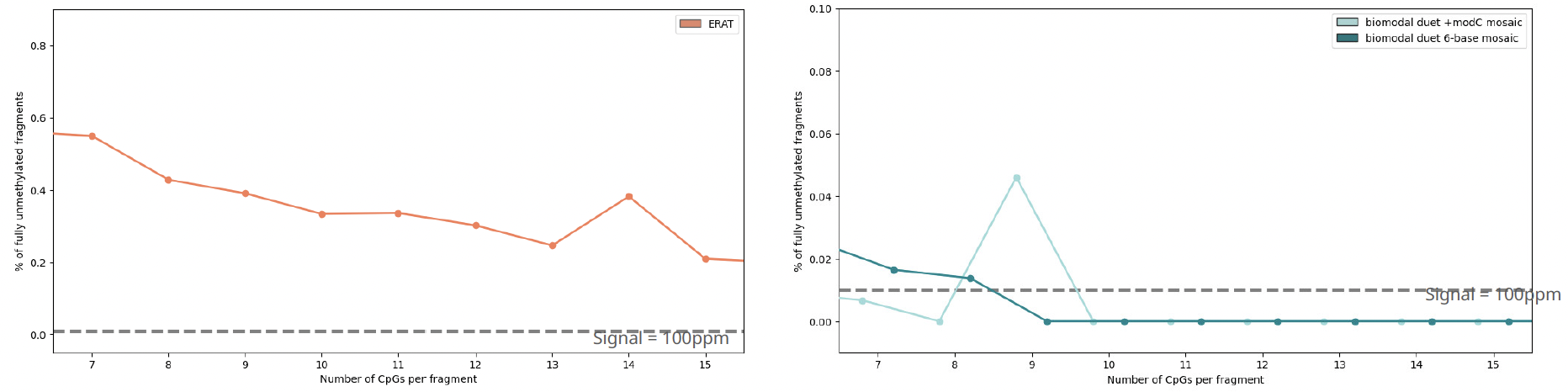
Fig. 5 ERAT-induced fragment hypomethylation sets a noise floor for ultra-low ctDNA detection. ctDNA concentration within cfDNA is <100ppm (0.01%)⁴. Within fragments with 7–15 CpG sites (typical for ctDNA molecules), we observed ~3,000ppm (0.3%) that appeared entirely hypomethylated when analysed with technologies that used ERAT during library preparation. This will impede the ability to detect genuine hypomethylation events within cfDNA samples. duet mosaic data contained hypomethylation artefacts below the 100ppm level.
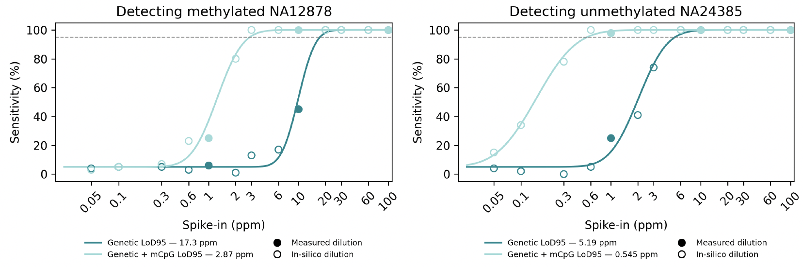
Fig. 6 duet 6-base mosaic LoD is improved through use of integrated genetic and methylation information from the same cfDNA fragment. We confirmed duet mosaic’s ability to achieve low LoD95 values by using Seraseq Unmethylated ctDNA Mutation Mix (NA24385) and methylated NA12878 alone or in spike in dilutions (1, 10 and 100 ppm). The two genomes differ at 6,366 homozygous SNVs, targeted using a 1Mb sequencing panel. Experimental dilutions were complemented by 4M-read in-silico mixtures at additional concentrations, generated by subsampling. Evidence for methylated NA12878 or unmethylated NA24385 was assessed using joint read-level methylation and SNV information or SNV information alone. LoD95 was estimated by probit regression (95% specificity). Combining genetic and epigenetic signal at single read-level enables an order of magnitude lower LoD95 compared to genetics alone, enhancing ctDNA detection capabilities.
duet mosaic workflows are optimised for cfDNA and use single stranded ligation to improve cfDNA capture, in particular of short fragments enriched in ctDNA⁵, and improve detection of 5mC and 5hmC. The elimination of ERAT further enhances liquid biopsy analyses through the removal of hypomethylation artifacts that confound the tissue of origin analysis required for MCED and CUP applications. Together, these improvements deliver limits of detection on the order of tens of ppm for single analytes, which can be improved to <1ppm when combined into read level genetic and epigenetic LoD.
The duet mosaic workflow meets the critical needs of liquid biopsy applications; complete and accurate phased multiomic features combined with recovery of the molecules that matter, resulting in ultra low LoD95 for ctDNA.
- Loyfer, N., Magenheim, J., Darwish, A., Isaac, S., Ganbat, J., Babikir, H., Jhutty, A., Wan, J., Bayés-Genís, A., Revuelta-López, E., Eden, A., Solanki, R., Dor, Y., & Kaplan, T. (2026). Systematic errors in enzymatic conversion limit cell-free DNA methylation specificity. bioRxiv. https://doi.org/10.64898/2026.03.24.713040
- https://www.illumina.com/science/genomics-research/articles/5-base-solution.html
- Loyfer, N., Magenheim, J., Peretz, A., Cann, G., Bredno, J., Klochendler, A., Fox-Fisher, I., Shabi-Porat, S., Hecht, M., Pelet, T., Moss, J., Drawshy, Z., Amini, H., Moradi, P., Nagaraju, S., Bauman, D., Shveiky, D., Porat, S., Dior, U., … Kaplan, T. (2023). A DNA methylation atlas of normal human cell types. Nature, 613(7943), 355–364.
- Melton CA, Freese P, Zhou Y, et al. A Novel Tissue-Free Method to Estimate Tumor-Derived Cell-Free DNA Quantity Using Tumor Methylation Patterns. Cancers (Basel). 2023;16(1):82. Published 2023 Dec 23. doi:10.3390/cancers16010082
- Mouliere, F. et al. Enhanced detection of circulating tumor DNA by fragment size analysis. Sci. Transl. Med. 10, eaat4921 (2018).